

Chronic kidney disease (CKD) is a common, globally significant condition, with associated significant mortality and morbidity. Globally, CKD has been ranked as the 18th most common cause of death, but with a disproportionately high loss of years of life associated with the condition (Jha et al, 2013). CKD is commonly classified according to glomerular filtration rate (GFR), with lower GFRs corresponding to worse disease, and a GFR of 5 representing ‘end-stage’ kidney disease (ESKD). For many advanced clinical practitioners (ACPs), CKD, dialysis and transplantation can be areas of clinical under-confidence due to their perceived specialist nature.
This article examines definitions of CKD, some key considerations in the clinical approach to a patient with CKD, including history and examination findings, and provides a brief overview of renal replacement strategies for the patient with ESKD. The article concludes with an overview of prescribing considerations for the patient with CKD.
Defining CKD
The following broad definition of CKD from the Kidney Disease Improving Global Outcomes group (KDIGO) has been widely adopted:
‘CKD is defined as abnormalities of kidney structure or function, present for >3 months, with implications for health.’
KDIGO, 2013: 5
This definition is included in UK guidance (National Institute for Health and Care Excellence (NICE), 2021a), and it is important to recognise that this is generally considered to be a progressive process. The rapidity of progression will vary between individuals, dependent upon underlying aetiology and comorbidity factors. This is different to a diagnosis of acute kidney injury (AKI) where there is an acute decline in kidney function. Although an in-depth consideration of AKI is beyond the scope of this article, it is recognised that AKI, especially when presenting on top of already present CKD, raises important safety issues, particularly with regard to medication management, so this is briefly addressed within the medication section.
The definition adopted by KDIGO reflects that patients may be classified as having CKD with a ‘normal’ GFR (>90 ml/minute (min)/1.73 m2). This may include: albuminuria (may be expressed as proteinuria); urine sediment abnormalities; electrolyte abnormalities due to tubular disorders; histological changes; structural change detected on imaging; or a history of kidney transplantation (KDIGO, 2013). Recognition of the potential for a diagnosis of CKD in the context of a ‘normal’ GFR is clinically important, as CKD confers a significantly increased risk of mortality and cardiovascular disease, even in patients without decline in their GFR (Matsushita et al, 2010).
Fulfilling criteria for a diagnosis of CKD does not indicate the cause of renal dysfunction, and patients will need further investigation to ensure the timely introduction of any therapies directed at optimisation of underlying pathological conditions. Key significant causes of CKD are listed in Table 1. Further detailed consideration of specific aetiologies is beyond the scope of this article, although diabetes, as the leading cause for CKD in the UK, is briefly reviewed in Box 1.
Table 1. Significant causes of chronic renal failure
| Disease | Approximate percentage of end-stage renal failure | |
|---|---|---|
| Diabetes | ~30% (reportedly higher incidence in the USA) | |
| Hypertension | Up to ~30% citedThough only represents 6.5% of incident RRT patients in the UK (2018 figures) | Incidence cited varies greatly |
| Glomerular diseases | 10-20% | eg nephropathy |
| Congenital/inherited disease | ~5% | eg Alport syndrome/polycystic kidney disease |
| Renal artery stenosis | ~5% | |
| Systemic inflammatory disease | ~5% | eg vasculitis/SLE |
| Unknown | Variable—up to 30% |
Key: RRT=renal replacement therapy; SLE= Systemic lupus erythematosus
Source: Turner et al, 2002; Vaidya and Aeddula, 2020; UK Renal Registry, 2020
Box 1.Diabetes and chronic kidney diseaseDiabetes is the leading cause of chronic kidney disease (CKD) worldwide. Approximately 40% of diabetic patients, of both type 1 and type 2 diabetes mellitus, develop diabetic kidney disease (DKD), referred to synonymously as diabetic nephropathy (DN). The aetiology of DKD is complex and multifactorial, but sustained hyperglycaemia and suboptimally controlled hypertension are the main causative factors. Genetic susceptibility, alongside glomerular hyperfiltration, smoking, obesity and physical inactivity are all known risk factors.The global incidence of DKD continues to increase, reflecting the increased prevalence of diabetes worldwide. A patient with longstanding diabetes of 10-plus years, the presence of albuminuria and a progressively reduced GFR results in a clinical diagnosis of DKD. Symptoms of DKD include fatigue, anorexia, and swelling of the extremities, although the condition is usually symptom free until disease is advanced. Treatment options include intensive control of hyperglycaemia, utilising a range of oral or injected medications, and management of hypertension with ACE inhibitors, angiotensin-receptor blockers (ARBs), or other antihypertensives. Lifestyle modification, including lipid reduction and smoking cessation may be beneficial for a patient with DKD.Source: Alicic et al, 2017; Leehey et al, 2021
Having defined the presence of CKD, it can be classified, according to GFR, within a five-stage classification promoted by KDIGO (2013). This classification represents a progressive worsening of kidney function, with stage 5 corresponding to ESKD. Stages of CKD are presented in Table 2. Notably, earlier stages of CKD are likely to be symptomless, although recognition of abnormal renal function is important in order to be able to monitor appropriately.
Table 2. Categorisation of chronic kidney disease
| Stage | GFR (ml/min/1.73 m2) | Description | Management |
|---|---|---|---|
| 1 | >90 | Normal kidney function maintained but urine or other abnormal findings suggest presence of disease | ObservationOptimisation of therapies to prevent progression |
| 2 | 60-89 | Mildly reduced function, urine or other abnormalities suggest presence of kidney disease | Secure diagnosisMonitoringBlood pressure control |
| 3a | 45-59 | Mildly to moderately decreased | As above—if not yet diagnosed, establish diagnosisOptimisation of medications/lifestyle factors |
| 3b | 30-44 | Moderately to severely decreased | |
| 4 | 15-29 | Severely decreased kidney function | Planning for ESRF/RRT |
| 5 | <15 | ESRF/established renal failure | Consideration of management of ESRF—conservative/HD/PD/transplantation |
Key: ESRF=end stage renal failure; GFR=glomerular filtration rate; HD=haemodialysis; PD=peritoneal dialysis; RRT=renal replacement therapy
Source: adapted from: KDIGO, 2013; Hunter, 2021; NICE, 2021aAlthough the terms ‘GFR’ and ‘eGFR’ (estimated GFR) are often used as synonymous, there may be variation in these measures, with GFR reflecting a direct measurement of renal function through one of several methods, and eGFR reflecting a derived, estimated measure of renal function based on measured serum creatinine. In clinical practice, eGFR is routinely used, as this is reported from most biochemistry laboratories as part of a readily available blood assay.
Although eGFR is routinely utilised, there are multiple potential pitfalls in interpretation of eGFR. Not least among these is the fact that multiple equations may be used for the calculation of eGFR in different settings, with significant variability. For example, Trevisani et al (2020) compared the accuracy of eight different equations, concluding that the degree of variance between eGFR and measured GFR increases as renal function deteriorates. Other sources note the development of more than 50 different equations to estimate eGFR (Luis-Lima and Porrini, 2017). Additionally, in extremes of body mass, or following intense physical activity or trauma, or when muscle mass is decreased (for example in anorexia or sarcopaenia), eGFR may be unrepresentative of true renal function (Rácz et al, 2012; Luis-Lima and Porrini, 2017). Such pitfalls or sources of error in eGFR estimation should be understood and taken into consideration in the clinical use of such measures (Thomas, 2019; UK Kidney Association, 2020).
Beyond staging of CKD according to eGFR, further classification of an individual's renal function according to level of albuminuria should be applied, again, as per KDIGO guidance (2013). Measurement of albumin:creatinine ratio (ACR) is advised for individuals with diabetes, and for those with a GFR of less than 60 ml/min/1.73 m2. Using ACR rather than protein:creatinine ratio (PCR) is suggested, as the latter is less sensitive for low levels of proteinuria (NICE, 2021a). Measurement of ACR is achieved through laboratory testing of urine samples, with figures of greater than 3mg/mmol considered clinically significant (NICE, 2021a).
Applying a quantification of albuminuria alongside GFR allows application of a framework supported by national guidance, with suggested frequency of follow-up and actions according to risk of progression of disease (see Table 3) (NICE, 2021a). Essentially, increasing albuminuria and decreasing GFR may both be considered markers of increased risk, with corresponding increasing levels of surveillance and follow-up suggested. UK guidance follows that of KDIGO, with NICE adopting the same criteria in its most recent clinical guideline (NICE, 2021a). A key message is that, while increased ACR and decreased eGFR both independently confer increased mortality, if present in combination there is a multiplicative effect on the risk of mortality (Matsushita et al, 2010; Kerr, 2012; NICE, 2021a).
Table 3. Suggested frequency of monitoring checks (of eGFR) per annum for adults, children and young people with, or at risk of, chronic kidney disease
| ACR category A1: normal to mildly increased (less than 3 mg/mmol) | ACR category A2: moderately increased (3 to 30 mg/mmol) | ACR category A3: severely increased (over 30 mg/mmol) | |
|---|---|---|---|
| GFR category G1: normal and high (90 ml/min/1.73 m2 or over) | 0 to 1 | 1 | 1 or more |
| GFR category G2: mild reduction related to normal range for a young adult (60-89 ml/min/1.73 m2) | 0 to 1 | 1 | 1 or more |
| GFR category G3a: mild to moderate reduction (45-59 ml/min/1.73 m2) | 1 | 1 | \2 |
| GFR category G3b: moderate to severe reduction (30-44 ml/min/1.73m2) | 1 to 2 | 2 | 2 or more |
| GFR category G4: severe reduction (15-29 ml/min/1.73m2) | 2 | 2 | 3 |
| GFR category G5: kidney failure (under 15 ml/min/1.73m2) | 4 | 4 or more | 4 or more |
Key: ACR=albumin creatinine ratio; GFR=glomerular filtration rate
Source: NICE, 2021aWithin England alone, there were around 1.8 million individuals with diagnosed CKD, in 2012, with an estimated further million undiagnosed (Kerr, 2012). Other studies, using self-reported measures, cite a prevalence of CKD in England of approximately 2%, similar for both men and women (NHS Digital, 2017). Both prevalence and severity of CKD increase with age, with 46% of over-75-year-olds having some degree of kidney disease (NHS Digital, 2017). The overwhelming majority of individuals diagnosed with CKD will be managed in a primary care setting. Notably, only a small percentage of patients diagnosed with CKD will go on to progress to ESKD, suggested at 1% and 20% of patients with stage 3 and stage 4 CKD respectively (Kshirsagar et al, 2008). However, even in the absence of disease progression, a significantly increased risk of death has been directly associated with a diagnosis of CKD (Kerr, 2012). One UK study found a 69% mortality at a mean follow-up period of 5.5 years, with 46% of deaths occurring from cardiovascular causes (Drey et al, 2003). Interestingly, and reinforcing the notion of limited progression to ESKD, only 4% of this cohort were established on a renal replacement therapy (RRT) by the end of follow-up. UK-wide figures support such limited progression to RRT. In 2018, 8000 new patients were commenced on some form of RRT (haemodialysis (HD)/peritoneal dialysis (PD)/transplant) and about 26 000 people were receiving HD or PD (UK Renal Registry, 2018). This suggests that <0.5% of diagnosed patients progress to RRT annually.
It should be recognised that, although UK-wide figures are cited above, the incidence and prevalence of CKD varies greatly depending on the population studied, including ethnic group and socio-economic class (Udayaraj, et al, 2013; Mathur et al, 2018).
Clinical assessment of the patient with CKD
Clinical findings from assessment of the patient with CKD will depend on the stage of disease experienced, with many patients having few or no symptoms, thus leading to late presentation (NICE, 2021a). In cases where symptoms are experienced, the clinical manifestation of renal disease has been referred to as ‘the uraemic syndrome’, reflecting the accumulation of urea, due to decreased renal clearance of this waste product—and this is one of the key drivers of symptoms (Vanholder et al, 2018; Dobre et al, 2019). Broadly speaking, patients at or approaching ESKD (CKD stage 5) can be expected to experience a greater burden of symptoms and are unlikely to be symptom free. Consideration of the overall impact of symptoms experienced is one of the key drivers to start RRT.
When examining a patient from a renal perspective, key questions to be answered relate to fluid status, and evidence of any coexistent pathology that may be contributing to renal dysfunction (for example hypertension). In the newly diagnosed patient with CKD, other findings in the history or examination may prove useful clues to guide further investigation, management and secure an underlying diagnosis. For example, a hearing aid user presenting with progressive CKD may prompt consideration of a diagnosis of Alport syndrome, a genetic disorder of the glomerular basement membrane, also affecting cochlear and ocular basement membranes, of variable genetic transmission and presenting commonly with haematuria as the first symptom (Kashtan, 2021). Similarly, knowledge of long-standing diabetes (or symptoms of the same) may support a diagnosis of diabetic nephropathy.
Multiple, non-specific symptoms such as, but not limited to, fatigue, nausea, decreased appetite, pruritus and spontaneous bruising are common in the patient with progressive CKD (Dobre et al, 2019). The insidious nature of symptom onset means they may not be mentioned by patients unless specifically explored. Hypertension will be present in a majority of patients with CKD (cited at 60-90% of patients dependent on stage and underlying aetiology of CKD) (Ku et al, 2019). Nocturia is a common early sign of CKD owing to a decreased ability to effectively concentrate urine in the ailing kidney. In patients with CKD, the prevalence of nocturia has been reported as being as high as 64%, with self-reporting of this symptom identified as an independent indicator of progression to ESKD (Lombardo et al, 2020). Fatigue, breathlessness or altered breathing patterns are common and may relate to a variety of causes, including renal anaemia, fluid accumulation or metabolic acidosis.
Cognitive symptoms
Patients presenting with advanced ESKD may demonstrate symptoms of uraemic encephalopathy, manifesting as confusion, agitation or other cognitive dysfunction, potentially with associated asterixis (a flapping tremor of the outstretched, dorsiflexed hands). It is likely to be seen in its more severe forms when eGFR falls below ~15 ml/min, although cognitive change may be evident with an eGFR in the 40-60 ml/min range (Olano et al, 2021). This phenomenon may be present in up to 60% of patients with CKD (Olano et al, 2021), and has been demonstrated in the majority of the haemodialysis population by some studies (Murray et al, 2006). If uraemia encephalopathy is excluded, alternate causes for these symptoms would need to be actively considered. These could include infection, hepatic encephalopathy, intra-cranial event, hypoglycaemia and hypertensive encephalopathy.
Anaemia symptoms
In the case of ESKD, patients will often appear generally unwell, with signs of anaemia and a pallid complexion (sometimes described as ‘lemon-yellow’) (Innes et al, 2018). As a result of CKD and associated uraemic accumulation, uraemic pruritus may be present. This results from a combination of factors, including drying of the skin, abnormal calcium and phosphorus metabolism, and general toxin accumulation (Vyas, 2010). On examination, scratches and excoriation may be evident, as may bruising, at least partly attributable to the effect on platelets of the uraemic milieu. CKD and particularly ESKD patients are at significantly increased risk of both bleeding and thrombotic complications due to a variety of complex pathophysiological interactions. Effective dialysis may partly mitigate bleeding risk. Lambert (2016) and Schrauben and Berns (2019) provide wider discussion on this topic. Although now rarely seen in a UK context, ‘uraemic frost’ on the skin is a marker of severity of disease (Madeux et al, 2016).
Skin symptoms
A cutaneous manifestation of renal disease that should not be missed is rash, potentially consistent with a vasculitic cause. This autoimmune condition may present cutaneously as a palpable, purpuric rash, which may, in the presence of deranged renal function, signal kidney involvement in the disease process. This will be confirmed by testing for specific antibodies (anti-neutrophil cytoplasm antibodies (ANCA)). A potential new diagnosis of ANCA vasculitis will generally involve referral to renal specialists for ongoing management. Further investigation will normally involve ultrasound assessment of the kidneys, and often progression to renal biopsy, with management dependent on the degree of renal impairment, chronicity of onset, and biopsy findings (McClure and Jones, 2018).
Hand, nail and arm symptoms
Examination of the hands and arms may demonstrate a variety of findings. Nail changes include leukonychia, Muehrcke's nails (bands of pale striations), Beau's lines, or ‘half and half’ nails, with lighter bands proximally (Neild et al, 2011). Patients should be assessed for splinter haemorrhages as potential markers for endocarditis. Assessing warmth and perfusion of both hands may provide useful information. For example, the presence of an arterio-venous (AV) fistula for dialysis access may cause diminished circulation (potentially vascular steal syndrome (Morris et al, 2020), noted in the ipsilateral hand as coolness or pallor. Conversely, hyperaemia due to increased flow may be present, with flushing and warmth of the ipsilateral hand in comparison to the non-fistula arm. Altered sensation may be consistent with peripheral neuropathy due to diabetic complications. Evidence of finger-prick testing will also indicate diabetes.
Hypertension
Blood pressure testing will commonly demonstrate hypertension in the newly diagnosed patient, or potentially normotension in patients maintained on anti-hypertensive agents. It is important to attempt to ascertain an individual patient's baseline blood pressure, from third-party sources if necessary, as patients may experience large shifts in blood pressure from baseline but still fall within ‘normal’ parameters (such as those outlined on early warning system documentation). As noted earlier, renal hypertension is very common (Ku et al, 2019) and, due to the complex and multifactorial nature of its pathophysiology, may be particularly refractory to treatment. It will therefore not be uncommon to find patients with CKD maintained on three or more pharmacological agents to manage hypertension. Renal hypertension is a significant topic of study in its own right, with multiple mechanisms contributing, including: overstimulation of the sympathetic nervous system; sodium retention; increased renin-angiotensin-aldosterone system (RAAS) activity; increased intracellular calcium levels and potential contributions from drugs such as erythopoetin administered to mediate the effects of renal anaemia (Ku et al, 2019). The difficulties are compounded by the fact that CKD contributes to the development of hypertension, while hypertension may also play a major role in development of CKD. Blood pressure control is thus key to slowing progression of CKD, with target systolic blood pressure of less than 140 mmHg advocated for adults with CKD and ACR <70 mg/mmol, and tighter control, with a target of less than 130 mmHg for adults with CKD and an ACR >70 mg/mmol (NICE, 2021a).
Due to the above factors, a multimodal approach to the management of renal hypertension is common, with drug treatments aimed at key targets in the contributing mechanisms outlined. UK guidance for patients with CKD (NICE, 2021a) is aligned to population-wide guidance (NICE, 2019), which promotes a step-wise approach to pharmaceutical management of hypertension, starting with an angiotensin-converting enzyme (ACE) inhibitor. Notably, patients with CKD and diabetes, or CKD and proteinuria >70 mg/mmol, even in the absence of hypertension, should also be offered an ACE inhibitor due to the potential improvement in proteinuria conferred by the medication. As discussed below, many medications need careful consideration in the CKD population, and ACE inhibitors are a good example of a medication needing careful introduction, due to potential deleterious effects on potassium regulation and renal function. Therefore, patients commenced on ACE inhibition, or having had a dose increase, should have bloods rechecked between 1 and 2 weeks from the change, in order to assess for this (NICE, 2021a). A decrease in eGFR of <25% of baseline, or increase in creatinine of <30% is acceptable according to national guidance (NICE, 2021b). Variation greater than these limits should prompt consideration of change of therapy and investigation for other potential contributing factors, including potential ‘unmasking’ of underlying reno-vascular disease (ie renal artery stenosis) (NICE, 2021b).
Arterio-venous fistula
Assessment of the arms may reveal the presence of an arteriovenous (AV) fistula, the accepted definitive choice of vascular access for the haemodialysis (HD) patient. This will normally be either radio-cephalic (radial artery to cephalic vein at the wrist), or brachio-cephalic/brachio-basilic at the elbow. The fistula is truly a lifeline for the HD patient, and they will generally be highly attuned to changes in its condition. Although multiple surgical sites of fistula formation are available, loss of options for HD access in the context of multiple failed fistulae may represent a treatment-limiting problem. Consequently, patients are trained not to allow any interventions to be performed on their fistula arm as these may jeopardise flow and patency. This includes blood pressure measurement, venepuncture and cannulation. Many HD patients will elect to wear an alert bracelet, silicone band or similar to alert first responders in the event of becoming unwell. For patients in hospital, an allergy band or similar should be applied to the fistula arm to reinforce this.
Fistulae should be assessed for pulsatile flow (a palpable thrill) and the presence or absence of a bruit through auscultation. Raising the arm while palpating will demonstrate any ‘collapsing’ of the fistula pulsation, supporting a diagnosis of hypovolaemia. Occasionally a fistula will develop to such an extent as to contribute to a heart failure symptomatology, and should be considered in the patient with heart failure symptoms (Stern and Klemmer, 2011). Referral for formal ultrasound assessment may be indicated in such situations, with surgical reduction of flow through the fistula potentially indicated.
Particularly in the context of systemic illness, decreased blood flow may lead to the failure of a fistula through thrombus formation. Dialysis patients will routinely check their own fistula and, if any concerns are raised, their first point of contact will normally be their renal unit for further investigation (ultrasound assessment as first investigation commonly) and consideration of alternate access to facilitate haemodialysis. A general assessment of the ESKD patient should also seek evidence of previous access formation attempts. This will be demonstrated through scarring consistent with alternate fistula locations and corroboration from the patient.
Elevated jugular venous pressure
Jugular venous pressure (JVP) is routinely examined as a marker of fluid status, reflecting right atrial pressure and thus used as a surrogate marker of the overall ‘filling’ status of the patient. If a patient is significantly fluid overloaded the practitioner may expect to find elevated JVP. As considered below, such an indication may trigger early consideration of initiation of dialysis in an oliguric ESKD patient. However, in interpreting JVP findings, it should be noted that there is a relative, perhaps surprising, dearth of literature in relation specifically to JVP in CKD. A small study of PD patients demonstrated poor correlation between clinical JVP findings and fluid overload in this patient group (Garfinkle and Barton, 2016). Regardless, assessment of the JVP remains an established component of a comprehensive renal examination.
For novice practitioners, it can be challenging to convincingly ascertain the JVP, so confirmation with an experienced practitioner may be useful until confident. The JVP is usually assessed with the patient reclined at 45 degrees to start, and the head turned away from the observer, but with the neck muscles relaxed. A normal (‘non-elevated’) JVP will be seen with point of maximal pulsation no more than 4 cm vertically above the sternal angle. Failure to see the JVP at 45 degrees should prompt progressively flattening or elevating the patient until JVP can be seen. Confirmation of JVP may be via a variety of techniques. It should be: impalpable (in comparison to carotid artery pulsation); occludable; demonstrate a double pulsation (assuming sinus rhythm); demonstrate respiratory variation in amplitude; and may be accentuated through firm pressure over the abdomen (abdomino-jugular reflux) (Innes et al, 2018; McGee, 2018).
Face and mucous membranes
Key facial signs to consider include anaemic changes such as pallor, which is best assessed in the mucous membranes of the lips and conjunctivae (Innes et al, 2018). If anaemia is suspected, this should be confirmed through testing of haemoglobin levels, then further investigated with regard to whether this reflects anaemia attributable to CKD. It is suggested that a haemoglobin level of less than 110g/litre, or symptoms attributable to anaemia, in a patient with an eGFR of <60 ml/min/1.73 m2 should prompt further investigation (NICE, 2021a). Investigations will normally include measurement of serum iron levels as well as ferritin and transferrin saturations, with interpretation of these results suggesting the best course of treatment. Common approaches to management of anaemia in CKD will include supplemental iron (if deficient), and commencement of erythropoietic-stimulating agents (ESA), followed by ongoing monitoring and dose titration (NICE, 2021a).
Assessment of mucous membranes is also included as a component of overall fluid assessment, with moistness or dryness being a somewhat subjective finding. The presence of xanthelasma may suggest hyper-lipidaemia as a modifiable contributor towards cardiovascular complications of CKD. Although uncommon in adults, facial oedema may be a marker of generalised hypoalbuminaemia, particularly in nephrotic syndrome (lower limb/dependent oedema being more common).
Heart, abdomen, lungs and lower limbs
Auscultation of heart sounds may reveal an ejection systolic murmur, audible throughout the praecordium, related to AV fistula flow. A third heart sound (‘S3’) may be heard and would add support to any suspicion of volume overload (Ramani and Weber, 2017). Of note, the ability to detect S3 on auscultation has been strongly correlated with the level of experience of the practitioner (Marcus et al, 2006). Uraemic pericarditis is a significant concern in the ESKD patient, and may be appreciated as a pericardial friction rub on auscultation. Left ventricular hypertrophy (LVH) is common in the chronically hypertensive patient, and may be suspected in the presence of a displaced apical beat or heaves. Assessment of the praecordium may demonstrate scars of previous line insertion sites. Many patients will have required temporary access, either emergently, or as a bridging measure to allow maturation of fistulae. Scars from either temporary central venous access catheters or tunnelled central venous catheters (a specialist dialysis catheter with longer potential dwell time) may be consistent with line placement to any of the large upper abdominal vessels, with subclavian and internal jugular sites perhaps being the most common. Finally, diminished heart sounds, elevated JVP, tachycardia and hypotension are potential markers of cardiac tamponade, a long recognised cardiac complication of the uraemic condition (Baldwin and Edwards, 1976; Bataille et al, 2015).
Examination of the chest may reveal, deep, sighing respirations (Kussmaul's breathing), as a compensatory response to the metabolic acidosis of CKD. Fluid accumulation and hypoalbuminaemia may lead to clinically appreciable pleural effusions and basal inspiratory crackles. While examining the posterior chest, it is customary to assess for any sacral fluid. This manifests in a dependent fashion as a ‘sacral pad’ with pitting oedema.
Abdominal examination follows the usual pattern, assessing visually for any asymmetry (potentially arising from large, polycystic kidneys or the presence of renal transplant graft in the left or right iliac fossa), or swellings. A suprapubic, smooth, central swelling may indicate a distended bladder, a potentially readily remediable cause of acute deterioration in function in CKD patients or patients presenting with new derangement in kidney function. The examiner will not be able to ‘get below’ a palpable bladder, and palpation will make discomfort worse in the patient with acute urinary retention. In this situation, urinary catheterisation will normally be indicated and should result in resolution of symptoms. Multiple scars may be noted, which may be prominent or subtle. Lower abdominal right or left Iliac fossa scars may correspond to previous renal transplantation, in which case a graft kidney should be palpable and non-tender in healthy states. Graft tenderness, either on palpation or spontaneously, warrants further investigation (normally via renal graft ultrasound in the first instance).
Native kidneys are not routinely removed for renal transplantation, but very large, or recurrently infective, or bleeding, polycystic kidneys may have been removed prior to or post-transplantation (or in non-transplant patients for similar indications). Increasingly, nephrectomy is performed laparoscopically, even for large polycystic kidneys, so laparoscopic surgery scars should prompt suspicion for potential nephrectomy (Eng et al, 2013). Open nephrectomy scars tend to be large, curvilinear and extend from flank to the lower costal margins of the posterior abdomen.
For patients undertaking PD a ‘Tenckhoff catheter’ will be present on the anterior abdominal wall, positioned to avoid interference with clothing and taking account of body habitus (physique). The abdominal wall exit site should routinely be inspected for any evidence of infection. Many ESKD patients may have trialled PD at some point, and perhaps been transitioned to HD, or received a transplant, in which case the catheter will have been removed, but small scars may be evident.
The technique of kidney palpation is best learnt by direct demonstration, but involves ‘ballotting’ of the kidneys between both hands, at the renal angles, with one hand anterior and one posterior, and the kidney gently mobilised back and forth between both hands.
Kidneys may not be palpable, dependent on body habitus, but if felt, they should be smooth, mobile and non-tender in healthy states. Renal angle tenderness on attempted palpation of kidneys is suspicious for ascending infection (pyelonephritis), stone disease, obstruction or abscess. Large, irregular kidneys may represent polycystic kidney disease, and a detailed family history may be helpful in corroboration. Auscultation of the abdominal vessels, and over the kidneys, may demonstrate bruits, suggesting turbulent flow and potential vascular disease, potentially supporting a differential diagnosis of reno-vascular disease (Ku et al, 2019).
Finally, assessment of the lower limbs may demonstrate pitting oedema, consistent with hypoalbuminaemia. This should be quantified by extent, eg ‘pitting oedema to knees’. Assessment of peripheral pulses, and exclusion of clinical signs of deep vein thrombosis, should also be undertaken. Peripheral neuropathy may be evident, dependent on underlying disease and comorbidity.
Renal replacement therapies
The appropriate timing of initiation of RRTs is a nuanced, individualised decision, in partnership between patients and their renal team. Commencement of the selected RRT modality is generally guided by the overall burden of symptoms. For some patients, signs and symptoms such as difficult-to-manage fluid retention, hyperkalaemia, or severe uraemia may trigger more rapid initiation. In some cases, symptoms may not prove troublesome, in which case an eGFR of 5-7 ml/min/1.73 m2 is considered a reasonable starting point for RRT (Tattersall et al, 2011; NICE, 2018).
Ideally, planning for RRT for CKD will have taken place in clinic settings prior to the need arising, based on the trajectory of decline of renal function, with national guidelines suggesting a year in advance of expected commencement (NICE, 2018). These discussions should focus on choice of modality of RRT as a patient-and-family-centred decision, guided by the renal team and with thought given to lifestyle factors and the individual patient's ability to manage aspects of RRT themselves. This planning phase should include consideration of access for RRT, either in the form of PD catheter insertion or arteriovenous fistula formation, with time allowed for healing and maturation of the fistula.
In ESKD the ‘gold standard’ of management may be considered renal transplantation, re-establishing normal or near-normal renal function and independence from supportive intermittent RRTs (Barclay and Burnapp, 2013). However, for a variety of reasons, ESKD patients may not be suitable candidates for transplantation (for example, they may have a high comorbidity burden, malignancy, be of advancing age, have psychosocial factors, or a very highly sensitised immunological status). Even if identified as a potential candidate for transplant (and the default position for referrers should be to assume ‘transplantability’ until proven otherwise, ensuring an informed discussion with patients prior to commencement on RRT (NICE, 2018)), the majority of patients will require a period of RRT in the interim. From a patient perspective, avoiding dialysis would be ideal, and as patients can be ‘activated’ on the transplant waiting list within 6 months of anticipated commencement of RRT, there is the possibility of ‘pre-emptive’ transplantation taking place (pre-dialysis). Where this does occur, it is often in the context of a live-donor transplant. The combination of live-donor transplantation to a pre-dialysis patient is likely to give the best possible outcome from the procedure. A detailed overview of renal transplantation is provided by Dunsmore (2019).
Alongside kidney transplantation, the two other main modalities of RRT available are PD or HD, and all options have advantages and disadvantages, summarised in Table 4. Additionally, in acute or acute-on-chronic kidney injury, continuous venovenous haemofiltration (CVVH) may be utilised, normally in an intensive therapy unit or high-dependency unit context. Although dialysis will maintain life, the toll that it takes on a patient's quality of life should not be underestimated, as recognised by Danovitch (2010):
‘… fatigue and malaise persist … Progressive cardiovascular disease, peripheral and autonomic neuropathy, bone disease and sexual dysfunction are common … Rehabilitation, particularly vocational rehabilitation, remains poor.’
Table 4. Comparison of modalities of renal replacement therapies
| Advantages | Disadvantages |
|---|---|
| Haemodialysis (HD) | |
|
|
| Peritoneal dialysis | |
|
|
| Renal transplantation (RT) | |
|
|
Adapted from Danovitch, 2010; Treharne et al, 2014; NICE, 2018; 2021a; Dunsmore, 2019
This is perhaps not surprising when one considers that even well dialysed patients will only achieve in the region of 15% of the normal waste clearance of two healthy kidneys (Danovitch, 2010).
It should also be recognised that, for some patients, declining dialysis and opting instead for a ‘conservative’ care approach may be a preferable option (sometimes referred to as a ‘non-dialytic approach’) (Noble et al, 2019).
To illustrate some key differences, Alston (2013) quotes the percentage of hospital attendance days at 4% versus 47% for conservative and HD approaches respectively, and also notes an average recovery time of 8 hours from each HD session. A conservative approach may be particularly worth consideration in patients of advancing age or with other comorbidities, which may make tolerating the demands of RRT challenging (for example severe cardiac or respiratory disease, dementia or cognitive decline). Of note, et al (2017) demonstrated equivalent self-reported quality-of-life measures between conservative care patients and those established on HD (although in a single centre study with a relatively small patient cohort). If a conservative approach is adopted, the patient will continue to be monitored by the renal team, with issues such as renal anaemia and mineral bone disorder still actively managed to optimise quality of life and independence, alongside close attention to management of symptoms arising from ESKD (Noble et al, 2019).
Haemodialysis
Intermittent HD remains the standard therapy for maintenance of metabolic stability in ESKD patients. Patients will attend hospital dialysis units up to three times weekly (many may opt for ‘self-care’ in a hospital setting, or home HD for a select cohort, both options after additional training from renal expert staff).
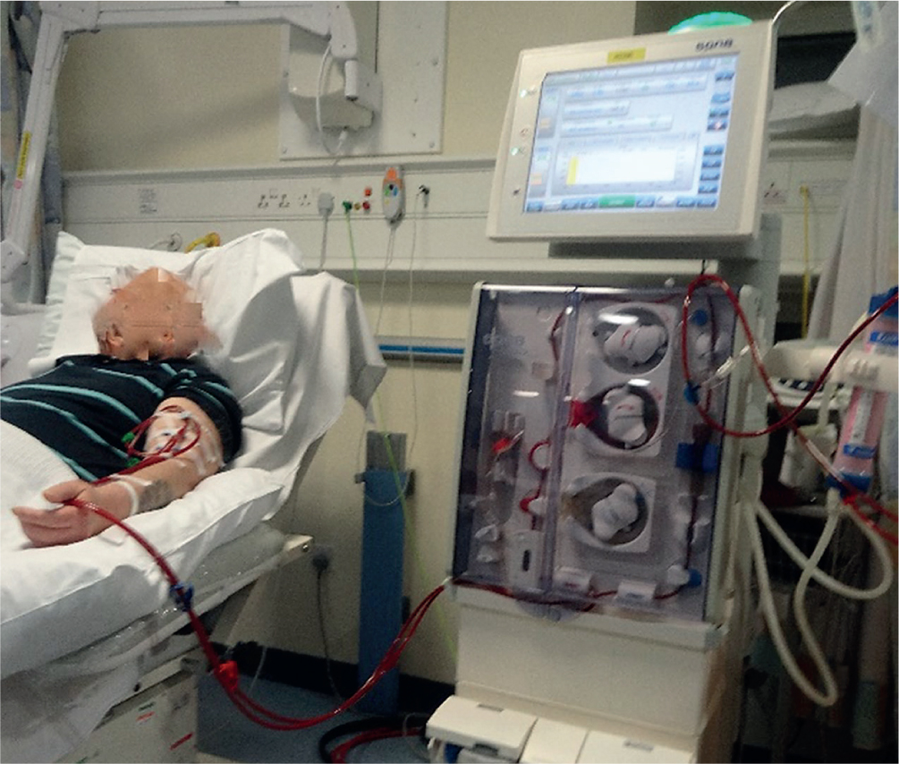
Having had a fistula formed in preparation, it will be ‘needled’ to allow placement of two large-bore cannulae, one nominally venous and one arterial, to allow flow of blood between the patient and the dialysis machine (Figure 1). HD works to extract waste products and excess fluid from the blood across a dialyser membrane, and uses a contraflow of the dialysate against blood flow to provide a concentration gradient.
An average dialysis session may last around 4 hours. The duration of individual sessions is dictated by regular and careful pre- and post-session blood sampling to determine the urea reduction ratio (URR), with most centres aiming to achieve a minimum ‘dose’ of 65% URR, and assessing this on a monthly basis. URR is used as a surrogate for a more complicated measure that relates to individual dialyser characteristics, the Kt/V (clearance, time/volume) for urea, (used to quantify dialysis treatment adequacy). Achieving higher URR is possible through longer treatment times, change of dialyser membrane, and changes to blood flow rates and dialysate flow rates (Hunter, 2021). Blood flow rate, and thus the efficiency of dialysis treatment, may be limited by type and adequacy of access and may prompt surgical revision of the fistula to improve this. Additional variables include the potassium and calcium concentrations of the dialysate fluid, bicarbonate concentration (to address the metabolic acidosis of CKD), dialysed temperature and choice of anti-coagulation.
Peritoneal dialysis
This form of dialysis requires insertion of a flexible dialysis catheter into the peritoneal space. The peritoneal membrane is then used as the ‘dialyser’ by instilling volumes of dialysis fluid into this potential space, via the PD catheter. Over the course of a ‘dwell time’, metabolic waste products diffuse from the peritoneal capillaries into the dialysis fluid. This fluid is drained off at the end of a cycle, and replaced with new, ‘clean’ dialysis fluid.
There are two main forms: continuous ambulatory peritoneal dialysis (CAPD) and automated peritoneal dialysis (APD). APD utilises a machine with automated warming and exchange functions and generally runs overnight, leaving patients free of dialysis during the day. CAPD involves a series of manual exchanges, usually ~4 times daily, but normal activity may be undertaken while dialysis is under way. In contrast to HD, PD is often viewed as a ‘gentler’ form of RRT, with less extreme shifts in fluid and biochemistry and may be a better fit for patients with underlying cardiovascular instability.
Economic considerations
If transplantation is undertaken, as well as the significant patient-centred improvements, there is also estimated to be a significant cost saving when compared to other modalities of RRT. A recent Swedish registry study estimates a €380 000 per patient saving over a 10-year period for transplantation compared with HD, even if patients ultimately return to HD due to graft failure (Jarl et al, 2018). Others have been more guarded in their analyses, with a recent systematic review suggesting diminishing savings from transplantation in those over 60 years of age (Fu et al, 2020).
Prescribing in kidney disease
Prescribing for the renal population can be challenging (Joint Formulary Committee, 2020). There are multiple classes of drugs that are particularly problematic, some discussed briefly below. When considering initiating any new medication in a patient with CKD, it will be wise to adopt a ‘start low, go slow’ approach to minimise risks of adverse reactions, accumulation or impact on renal function. Increased frequency of monitoring may be a reasonable safety-netting procedure.
A low threshold should be adopted for discussion with renal specialist services and particularly with renal specialist pharmacists if there is any ambiguity. An important reference resource, readily to hand in any renal unit, is the Renal Drug Handbook (Ashley and Dunleavy, 2018). Additionally, in the RRT population, consideration needs to be given to the potential dialysability of medications (ie is the medication removed by dialysis), as this will impact timing of doses. For ACPs, knowledge of common drugs to avoid or prescribe with significant caution is crucial.
Examples of key medications and concerns for ACPs
Antimicrobials
The following antimicrobials are problematic for patients with renal problems.
- Nitrofurantoin is not recommended for patients with an eGFR <60 ml/min/1.73 m2, due to risks of peripheral neuropathy and lack of effectiveness in treatment of urinary tract infections due to poor urine concentration
- Gentamicin is generally avoided in acute kidney injury, and in acute-on-chronic kidney injury. It may be used in patients with advanced stages of CKD, but this will be via specific protocols
- Macrolide antibiotics may have a particularly potent effect on elevating serum levels of certain immunosuppressant medications (for example in the post-transplant patient), these interactions may drive nephrotoxicity and impact on function in this group
- Protocols for therapeutic drug monitoring of antibiotics may also be quite different in patients with ESKD, for example regarding vancomycin.
Non-steroidal anti-inflammatory drugs
Non-steroidal anti-inflammatory drugs (NSAIDs) should be avoided if possible in all levels of renal dysfunction. Prolonged use may decrease native urine output irreversibly in patients on RRT. Chronic usage is associated with progression of CKD, and acute use with a decrease in eGFR (normally reversible) (NICE, 2021a). Use may be appropriate in particular patients if there are no good alternatives, and following discussion.
Opioids
Opioids are generally not advised in CKD, due to the potential for accumulation and significant adverse effects. Formulations considered at lower risk of accumulation include fentanyl and alfentanil, but these should be introduced at a low dose of an immediate release preparation and actively monitored for side effects. Avoidance of modified release formulations of opiates has been recommended. (Joint Formulary Committee, 2020; Whitworth and Thomson, 2021).
Diuretics and antihypertensives
The optimal approach to the management of diuretic and antihypertensive medication will depend on the level of CKD and if there is any acute deterioration in kidney function. It should be recognised that patients with CKD are part of a wider group in whom these medications present particular risks of avoidable harm. Patients admitted to hospital or experiencing episodes of an acute intercurrent illness need particularly careful consideration.
The ‘Think Kidneys’ initiative has provided key guidance on a pragmatic approach to the stopping and restarting of these high-risk medications (Think Kidneys, 2020), which has been widely adopted. It includes the use of ‘Sick Day Rules’ to assist patients in optimising their own care.
A shared care approach to managing medication changes should be adopted wherever possible. Discussion with patients is a key factor in decision-making around stopping, withholding and restarting medications and the attendant risks and anticipated benefits of each potential course of action.
Conclusion
CKD represents a broad spectrum of disease, from asymptomatic abnormalities of biochemistry, through to multi-system manifestations of kidney disease, requiring intensive, three-times-weekly technological interventions to maintain life. CKD is common, and confers a high burden of morbidity and mortality. The ACP has a significant role to play regardless of the context of their practice. The majority of patients with CKD are managed in a primary care setting, where close attention to optimising underlying conditions and mitigating the effects of renal dysfunction are key to ensuring better outcomes. In secondary care, patients with CKD may present with a variety of related complications, or novel pathology unrelated to CKD, but complicated by its presence. These patients all require a nuanced, multidisciplinary approach in order to optimise their outcomes. Prescribing for this group may be challenging and close liaison with specialist services is recommended.
KEY POINTS
- Chronic kidney disease (CKD) is a common condition which the majority of advanced clinical practitioners (ACPs) will encounter in clinical practice
- The role of the ACP in primary or secondary care may focus on identifying the presence of CKD or preventing further decline
- Progressive renal dysfunction as a patient progresses through stages of CKD confers high morbidity and burden of symptoms, and significantly increases cardiovascular risk
- Differing modalities of renal replacement therapy have particular advantages and challenges, and the ‘correct’ choice is an individualised decision
- Prescribing for a patient with CKD is complex, with dosing affected by reduced kidney function or renal replacement therapies
- Care of the CKD patient is a complex, interdisciplinary undertaking, with multiple professional groups playing key roles
CPD reflective questions
- Reflect on common medication prescriptions which require caution for patients with chronic kidney disease (CKD). How would you ensure safety if patients in your practice area required these medications?
- Consider how to support a CKD patient who comes under your care, particularly in relation to end-of-life care
- What are the key elements within a person-centred approach when consulting with, and caring for, a patient with chronic kidney disease?