References

Cantini F, Niccoli L, Storri L Are polymyalgia rheumatica and giant cell arteritis the same disease?. Semin Arthritis Rheum.. 2004; 33:(5)294-301 https://doi.org/10.1016/j.semarthrit.2003.09.008

Chu R, Foster C, Ali M Optimal length and usefulness of temporal artery biopsies in the diagnosis of giant cell arteritis: a 10-year retrospective review of medical records. Lancet Rheumatology.. 2020; 2:(12)E774-E778 https://doi.org/10.1016/S2665-9913(20)30222-8

Cummings GG, Fraser K, Tarlier DS. Implementing advanced nurse practitioner roles in acute care: an evaluation of organizational change. J Nurs Adm.. 2003; 33:(3)139-145 https://doi.org/10.1097/00005110-200303000-00004

Dasgupta B Concise guidance: diagnosis and management of giant cell arteritis. Clin Med (Lond).. 2010; 10:(4)381-6 https://doi.org/10.7861/clinmedicine.10-4-381

Dasgupta B, Borg FA, Hassan N BSR and BHPR guidelines for the management of giant cell arteritis. Rheumatology (Oxford).. 2010; 49:(8)1594-1597 https://doi.org/10.1093/rheumatology/keq039a

Polymyalgia rheumatica and giant cell arteritis. In: Dasgupta B, Dejaco C (eds). Oxford: Oxford Rheumatology Library/Oxford University Press;

Dejaco C, Duftner C, Buttgereit F, Matteson EL, Dasgupta B. The spectrum of giant cell arteritis and polymyalgia rheumatica: revisiting the concept of the disease. Rheumatology (Oxford).. 2017; 56:(4)506-515 https://doi.org/10.1093/rheumatology/kew273

There's a S.M.A.R.T. way to write management's goals and objectives. 1981. https://tinyurl.com/yvdmj4kv (accessed 21 April 2021)

González-Gay MA, García-Porrúa C. Epidemiology of the vasculitides. Rheum Dis Clin North Am.. 2001; 27:(4)729-749 https://doi.org/10.1016/s0889-857x(05)70232-5

González-Gay MA, Vazquez-Rodriguez TR, Lopez-Diaz MJ Epidemiology of giant cell arteritis and polymyalgia rheumatica. Arthritis Rheum.. 2009; 61:(10)1454-1461 https://doi.org/10.1002/art.24459

Hellmich B, Agueda A, Monti S 2018 update of the EULAR recommendations for the management of large vessel vasculitis. Ann Rheum Dis.. 2020; 79:(1)19-30 https://doi.org/10.1136/annrheumdis-2019-215672

Mackie SL, Dejaco C, Appenzeller S British Society for Rheumatology guideline on diagnosis and treatment of giant cell arteritis. Rheumatology (Oxford).. 2020; 59:(3)e1-e23 https://doi.org/10.1093/rheumatology/kez672

Mackie SL, Brouwer E. What can negative temporal artery biopsies tell us?. Rheumatology (Oxford).. 2020; 59:(5)925-927 https://doi.org/10.1093/rheumatology/kez628

Mollan SP, Sharrack N, Burdon MA, Denniston AK. Aspirin as adjunctive treatment for giant cell arteritis. Cochrane Database Syst Rev.. 2014; 8 https://doi.org/10.1002/14651858.CD010453.pub2

Mollan SP, Paemeleire K, Versijpt J, Luqmani R, Sinclair AJ. European Headache Federation recommendations for neurologists managing giant cell arteritis. J Headache Pain.. 2020; 21:(1) https://doi.org/10.1186/s10194-020-01093-7

National Institute for Clinical Excellence. Giant cell arteritis. Clinical knowledge summary. 2020. https://cks.nice.org.uk/giant-cell-arteritis (accessed 21 April 2021)

National Institute for Clinical Excellence. Tocilizumab for treating giant cell arteritis. Technology appraisal guidance: TA518. 2018. https://www.nice.org.uk/guidance/ta518 (accessed 21 April 2021)

National Institute of Arthritis and Musculoskeletal and Skin Diseases. Polymyalgia rheumatica and giant cell arteritis. 2016. http://www.niams.nih.gov/Health_Info/Polymyalgia/default.asp (accessed 21 April 2021)

National Organisation of Rare Diseases. Giant cell arteritis. 2019. https://rarediseases.org/rare-diseases/arteritis-giant-cell/ (accessed 21 April 2021)

Neville L, Swift J. Measuring the impact of the advanced practitioner role: a practical approach. J Nurs Manag.. 2012; 20:(3)382-9 https://doi.org/10.1111/j.1365-2834.2012.01356.x

The role of the advanced nurse practitioner. Blog. 2019. https://www.independentnurse.co.uk/blogs-article/the-role-of-the-advanced-nurse-practitioner/213167/ (accessed 21 April 2021)

Enduring ambiguity: the ANP role. 2020. https://www.nursinginpractice.com/professional/enduring-ambiguity-the-anp-role/ (accessed 21 April 2021)

Poller DN, van Wyk Q, Jeffrey MJ. The importance of skip lesions in temporal arteritis. J Clin Pathol.. 2000; 53:(2)137-9 https://doi.org/10.1136/jcp.53.2.137

Royal College of Nursing. Royal College of Nursing standards for advanced level nursing practice. 2018. https://www.rcn.org.uk/professional-development/publications/pub-007038 (accessed 21 April 2021)

Rubenstein E, Maldini C, Gonzalez-Chiappe S, Chevret S, Mahr A. Sensitivity of temporal artery biopsy in the diagnosis of giant cell arteritis: a systematic literature review and meta-analysis. Rheumatology (Oxford).. 2020; 59:(5)1011-1020 https://doi.org/10.1093/rheumatology/kez385

Sait M, Lepore M, Kwasnicki R The 2016 revised ACR criteria for diagnosis of giant cell arteritis—our case series: can this avoid unnecessary temporal artery biopsies?. International Journal of Surgery Open.. 2017; 17:19-23 https://doi.org/10.1016/j.ijso.2017.09.003

Salvarani C, Cantini F, Hunder GG. Polymyalgia rheumatica and giant-cell arteritis. Lancet.. 20081; 372:(9634)234-45 https://doi.org/10.1016/S0140-6736(08)61077-6

Giant cell arteritis (temporal arteritis) workup. 2020. https://emedicine.medscape.com/article/332483-workup#c7

Sharma A, Mohammad AJ, Turesson C. Incidence and prevalence of giant cell arteritis and polymyalgia rheumatica: a systematic literature review. Semin Arthritis Rheum.. 2020; 50:(5)1040-1048 https://doi.org/10.1016/j.semarthrit.2020.07.005

Smeeth L, Cook C, Hall AJ. Incidence of diagnosed polymyalgia rheumatica and temporal arteritis in the United Kingdom, 1990-2001. Ann Rheum Dis.. 2006; 65:(8)1093-1098 https://doi.org/10.1136/ard.2005.046912

Tuckwell K, Collinson N, Dimonaco S Newly diagnosed vs. relapsing giant cell arteritis: Baseline data from the GiACTA trial. Semin Arthritis Rheum.. 2017; 46:(5)657-664 https://doi.org/10.1016/j.semarthrit.2016.11.002

Vasculitis UK. What is giant cell arteritis/temporal arteritis?. 2019. https://tinyurl.com/45sk627y (accessed 21 April 2021)

BMJ best practice. Giant cell arteritis. 2020. https://bestpractice.bmj.com/topics/en-gb/177 (accessed 21 April 2021)

Weyand CM, Liao YJ, Goronzy JJ. The immunopathology of giant cell arteritis: diagnostic and therapeutic implications. J Neuroophthalmol.. 2012; 32:(3)259-65 https://doi.org/10.1097/WNO.0b013e318268aa9b

Wong CA, Cummings GG, Ducharme L. The relationship between nursing leadership and patient outcomes: a systematic review update. J Nurs Manag.. 2013; 21:(5)709-724 https://doi.org/10.1111/jonm.12116

Yates M, Graham K, Watts RA, MacGregor AJ. The prevalence of giant cell arteritis and polymyalgia rheumatica in a UK primary care population. BMC Musculoskelet Disord.. 2016; 17 https://doi.org/10.1186/s12891-016-1127-3

Introducing an ANP-led temporal artery biopsy service for patients with suspected giant cell arteritis

13 May 2021
Volume 30 · Issue 9

Abstract

Giant cell arteritis (GCA) is an uncommon autoimmune inflammatory vasculopathy that can lead to the destruction and occlusion of various arteries that consequently can cause serious complications such as stroke or sight loss. It is seen as a medical emergency. The most commonly affected vessel in GCA is the temporal artery in the side of the head, hence the condition is sometimes also referred to as ‘temporal arteritis’. This article discusses the introduction of an advanced nurse practitioner-led temporal artery biopsy service.

This article provides a summary of the symptoms, diagnosis, treatment and management of giant cell arteritis (GCA) in order to provide context for a review of the introduction of an advanced nurse practitioner (ANP)-led temporal artery biopsy service (TAB).

According to the National Institute for Health and Care Excellence (NICE) (2020), GCA is defined as an autoimmune chronic vasculitis which is associated with inflammation within the muscular walls of the arteries. The condition is sometimes also referred to as ‘temporal arteritis’ due to the temporal artery in the side of the head often being affected (Vasculitis UK, 2019) (Figure 1).

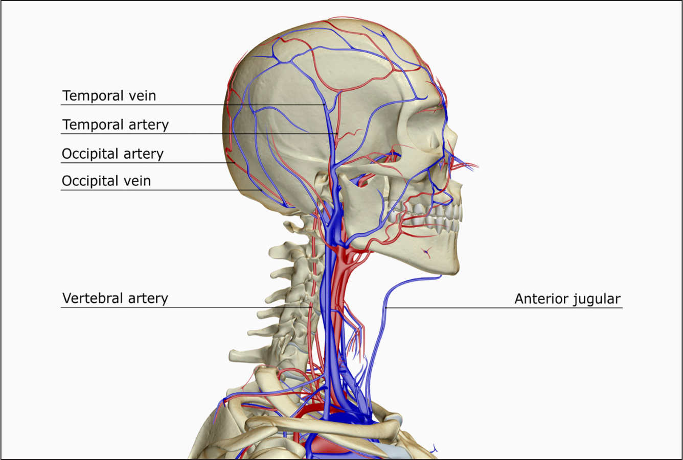
Figure 1. A lateral view of the blood supply of the head and neck, showing the position of the temporal artery

The cause of GCA is unknown, although genetic and environmental factors have been linked to a predisposition to the disease (Genetic and Rare Diseases Information Center (GARD), 2017). GCA is predominately associated with affecting the medium to large arteries in the head, especially extracranial branches of the carotid artery and the ophthalmic artery (NICE, 2020). Other arteries elsewhere in the body can also be involved, including, for example, the aorta, but this is uncommon within those diagnosed with GCA (National Institute of Arthritis and Musculoskeletal and Skin Diseases, 2016).

The destructive inflammatory process associated with GCA causes the internal lumen of the artery to narrow (luminal stenosis) and even to completely occlude, potentially contributing to vascular insufficiency and ischaemia in the adjacent and associated tissue.

The immunopathology of GCA specifically relates to the production of granulomatous infiltrates (a collection of immune cells called macrophages whose function is to ingest bacteria). These provoke a cellular ‘response to injury’ or amplification cascade that, in turn, causes vascular injury and hyperplasia (enlargement of tissue) within the walls of the arteries, leading to their potential remodelling, destruction and luminal narrowing (Weyand et al, 2012; Dasgupta and Dejaco, 2016).

Obstruction of arterial blood supply to the eye (via the ophthalmic artery) as a consequence of luminal stenosis caused by GCA can lead to a reduction in, or an acute loss of, vision.

GCA is rare and research findings vary as to the exact extent and distribution of the disease, but it is estimated that the number of people affected in the UK is 17–22 per 100 000 (Yates et al, 2016; Sharma et al, 2020, NICE, 2020). It is 2-3 times more common in females and 7 times more common within the white Caucasian population (González-Gay, 2001). GCA in Asian, African American and Hispanic populations is much less significant (Dasgupta and Dejaco, 2016). GCA is more prolific in the Scandinavian population and in parts of North America (Sharma et al, 2020) and there is an increased geographical prevalence in northern compared to southern Europe (González-Gay et al, 2009). GCA is more prevalent in individuals over the age of 50, and particularly in those aged 70-79 years (Smeeth, 2006; NICE, 2020).

There is a strong correlation between GCA and another autoimmune inflammatory disorder known as polymyalgia rheumatica (PMR). About 50% of patients with confirmed GCA will also have PMR (Cantini et al, 2004; Dejaco et al, 2017).

Symptoms of GCA

GCA is associated with several potentially insidious symptoms; however, none of them are specific enough (pathognomonic) to the disease to be definitively diagnostic. Typically, patients will present with a new-onset unilateral localised temporal headache that occasionally can be bilateral and more diffuse (Salvarani et al, 2008). Tuckwell et al (2017) reported that new onset headache is the most commonly reported symptom of GCA, reflected in 67% of patients.

As many as 20% of patients present with visual symptoms (Dasgupta and Dejaco, 2016). This can constitute a partial or even complete loss of vision. This is due to compromised blood supply to the optic nerve head (known as anterior ischaemic optic neuropathy (AION)) or within the retina (central retinal artery occlusion (CRAO)). Some patients experience brief intermittent transient loss of vision (known as amaurosis fugax), which may proceed complete loss of vision and represents an important visual symptom. Other visual symptoms may include double vision (diplopia), or visual field defects. Fundoscopy (examination of the retina) may elicit a pale oedematous optic disc, ‘cotton wool’ spots and haemorrhages in the retina suggestive of vascular deficiency. Usually, if the patient presents with visual symptoms, they are likely to be unilateral. However, despite an initial unilateral presentation, both eyes can be affected within 1 to 2 weeks if left untreated, which prompts urgent treatment in order to prevent this. Often any significant visual loss prior to treatment is permanent and irreversible, particularly in older age groups (Dasgupta and Dejaco, 2016).

The temporal artery is often affected by the disease process and if so, can be tender and painful to touch. This discomfort may also be reflected to the scalp area—some patients may complain of pain when brushing their hair, for example. The temporal artery can also become thickened and enlarged, causing the pulse to become reduced or even absent. Conversely, in some patients, the artery can externally be quite visible, having a tortuous and pronounced route as it transverses the forehead.

Another classic symptom that affects approximately half of all GCA-positive patients is intermittent jaw ache or pain when eating, known as jaw claudication. This is due to ischaemia of the masticatory muscles (Salvarani et al, 2008). There can also be other less specific and generic symptoms such as low-grade fever, neurological features, weight loss, fatigue, peripheral oedema, cough, sore throat and depression (National Organisation of Rare Diseases, 2019).

Diagnosis of GCA

Serum blood tests

The patient with suspected GCA will often attend the acute services with some of the symptoms outlined above. Initially, a thorough clinical history will be required, which may provide some additional clues, such as the patient having PMR, for example. Along with a patient history, a series of serum blood tests and more involved investigations (ultrasound imaging and TAB) may be considered to enable diagnosis. There are several potential differential diagnoses and these are outlined in Box 1.

Box 1.Other potential differential diagnoses for GCA

  • Herpes zoster (shingles)
  • Cluster headache or migraine
  • Acute angle closure glaucoma
  • Retinal transient ischaemic attack
  • Temporomandibular joint pain
  • Sinus disease and ear problems
  • Cervical spondylosis or other upper cervical spine disease
  • Ankylosing spondylitis
  • Cranial malignancy
  • Intracranial pathology

Source: NICE, 2020

The British Society of Rheumatology guidelines (Mackie et al, 2020) suggest that, once there is a suspicion of GCA, routine blood tests may be useful to support the diagnosis including:

  • Erythrocyte sedimentation rate (ESR)
  • C-reactive protein (CRP)
  • Platelet count (as part of full blood count)
  • Liver function test (LFT): alkaline phosphatase specifically
  • Baseline plasma glucose.

ESR and CRP are non-specific laboratory markers of inflammation, both of which can be raised in GCA. Typically, an ESR of >50 mm/hour and a CRP >20 mg/litre can be regarded as being elevated and may be suggestive of GCA (Warrington, 2020). However, there has to be a degree of caution in assuming that raised serum blood results are conclusively suggestive of GCA as there can be other underlying reasons that may cause inflammatory markers to be raised, such as anaemia, pregnancy, diabetes mellitus, infection and malignancy.

Among other related serum blood tests, a raised platelet count (>300 x 109/litre) in correlation with raised inflammatory markers may also be diagnostic of GCA. Tests for antinuclear antibodies, antineutrophil cytoplasmic antibodies, glucose, creatinine, bone profile and thyroid function may be undertaken in order to rule out other potential clinical diagnoses.

Ultrasonography vs temporal artery biopsy

Colour-duplex ultrasonography (CDUS) is a non-invasive imaging test that may be used to diagnose GCA. The spontaneous on-screen colour imaging ultrasound test allows the user to assess the thickness and lumen of the artery wall. In a GCA-positive ultrasound test a cross-section of the artery may show a ‘halo sign’, which is a hypoechogenic (darkening) signal highlighted between the artery layers (lamina).

Previously, the role of imaging techniques in the diagnosis of GCA has been undervalued as it was argued that the sensitivity of this test was lower than that of the TAB procedure (Mollan et al, 2020). However, Mackie et al (2020) noted that a meta-analysis of recent studies implies that ultrasound is more sensitive but less specific than TAB and has a particular benefit in ruling out low probability or confirming high probability cases. Statistical measures of test performance, sensitivity (true positive test) and specificity (true negative test) enable clinicians to contextualise test accuracy. In relation to the diagnosis of GCA, comparison of the results of TABs verses CDUS as viable tests to determine diagnosis continue to cause some debate in the literature (Mackie and Brouwer, 2020). For example, another very recent meta-analysis stated that the sensitivity of TABs is 77% compared to 55–77% for CDUS (Rubenstein et al, 2020). However, it is argued that these figures are to some extent reliant on various extraneous factors, such as the experience of the sonographer in undertaking CDUS (Mackie and Brouwer, 2020).

Temporal artery biopsy

Previously, TAB had been widely recognised as the gold-standard procedure in the confirmation of GCA (Dasgupta and Dejaco, 2016). Recent evidence is more equivocal and, given the more accepted role of imaging, it may be undertaken either in conjunction with or instead of a TAB. Ideally, confirmation of the diagnosis with both ultrasound and a TAB would be preferable, but not always practically possible (Mackie et al, 2020).

It is worth noting that due to the rare occurrence of ‘skip lesions’ the section of artery harvested during a TAB may not be representative of the rest of the vessel. This can lead to a negative result despite the presence of GCA. This is known as a false-negative, and in study by Poller et al (2000) this represented a mere 8.5% of patients with active vasculitis. However, this does show that TABs have their shortcomings and the possibility of this potential outcome needs to be explained to the patient during the consent process.

The TAB is an invasive surgical procedure that involves harvesting a short section of the temporal artery on the side of the head that is associated with displaying most of the symptoms. Usually the procedure is undertaken in a theatre suite under a local anaesthetic.

A TAB procedure initially involves the trajectory of the artery being marked out on the skin (the hair may have to be removed in the vicinity) of the temple. If the artery is difficult to palpate or visualise then a hand-held Doppler ultrasound can be used to locate the vessel. Once identified and marked, a subcutaneous local anaesthetic is administered into the area.

The skin is cleaned and draped for surgery. An incision is then made through the skin of approximately 4 cm in length, either along or adjacent to the vessel. Soft-tissue dissection is undertaken through the dermis and underlying fat to expose the superficial fascia and the artery. Once identified, a length of artery will be resected from the surrounding tissue, isolated at each end and ligated with suture. Then a section (approximately 15-20 mm) between the ligated ends is excised and transferred to a histopathology pot and sent to the laboratory. The wound will be checked to ensure that there is no bleeding and then stitched back together. The procedure usually takes approximately 30 minutes, depending on how easy the artery is to locate.

As with any invasive procedure a TAB is associated with surgical risks, including bleeding, swelling, infection and wound dehiscence, but is generally regarded as a safe procedure (Dasgupta and Dejaco, 2016). In the very rare circumstance where the normal anatomy is altered and the external carotid artery contributes to the collateral circulation to the brain (ordinarily, the brain receives its blood supply from the two internal carotid and vertebral arteries via the Circle of Willis) there may be a slightly increased risk of a ischaemic stroke as a consequence of the procedure (Seetharaman et al, 2020). Brow ptosis (droop) may occur if the facial nerve is damaged during the procedure. Although these risks are extremely rare, careful consideration is required by the patient during the consenting process.

Once the biopsy has been histologically processed, the result will either be positive or negative for GCA. It may also show other aspects of inflammation or damage.

Treatment and management of GCA

If a patient attends with symptoms strongly suggestive of GCA, guidance (Mackie et al, 2020) advises that they should be immediately started on high-dose glucocorticoid (usually prednisolone 60 mg) until such time as a more definitive diagnosis can be elicited. High-dose glucocorticoids are associated with several undesirable side-effects, but over a short period (until diagnosis) the risk is low. Long-term risks include weight gain, hypertension, diabetes, mood swings, osteoporosis and fluid retention. Any plasma blood tests such ESR, CRP and platelets being undertaken in relation to diagnosis should ideally be carried out before starting on glucocorticoids, as these drugs may affect the results.

NICE guidance suggests that a CDUS and/or TAB should be ordered to confirm the suspicion of GCA. The patient should then be referred either to a rheumatologist or, if there are eye-related symptoms, an ophthalmologist (or both) the same day (NICE, 2020).

Once started on oral glucocorticoids, the clinical response should be monitored for the first few days and to also check if there have been any undesirable side effects due to the steroids.

Patients with acute visual loss may initially be given intravenous methylprednisolone 500–1000 mg once a day for 3 days and then started on oral glucocorticoids.

Once GCA has been confirmed, the patient is likely to stay on prednisolone for 12 to 18 months, provided there are no significant side effects. Multidisciplinary monitoring will be required during the treatment period and the dose of steroids is likely to be tapered down over time. Along with polymyalgia, GCA accounts for one of the most common reasons for long-term steroid treatment in the community (Desgupta and Dejaco, 2016).

Calcium and vitamin D supplements are also prescribed during the glucocorticoid treatment due to the risk of osteoporosis caused by inhibition of calcium absorption and suppression of bone development instigated by the steroids. Some clinicians also advocate the use of aspirin in order to reduce the auto-immune effects of the disease and the potential damage to the artery walls; however, this remains controversial (Mollan et al, 2014).

Alternative treatments to glucocorticoids include disease-modifying anti-rheumatics including methotrexate and, more recently, biological therapy (tocilizumab) (NICE, 2018).

Once steroid treatment is started, and a TAB is indicated, there is a 14-day window of opportunity to undertake the procedure as the steroid treatment will reduce the inflammatory response and can subsequently influence the result (Dasgupta, 2010). Moreover, a recent study by Sait et al (2017) suggested that the diagnostic yield for a TAB may begin the wane in as little as 8 days following commencement of steroids. Conversely, there is suggestion that a TAB result is still viable up to 6 weeks after starting steroids, but delaying that long is rarely recommended (Dasgupta et al, 2010).

The ANP-led TAB service

Advanced nurse practitioner roles have developed significantly in the UK over the past 20 to 30 years and recently have been described as ‘invaluable’ in co-ordinating timely patient care and negating unnecessary treatment delay (Osho, 2019). Many complementary ANP roles within both primary and tertiary care have been identified that work collaboratively within the multidisciplinary team across an abundance of specialties including ophthalmology. The Royal College of Nursing (2018) described the ANP as a registered nurse who, among other things, undertakes autonomous decision-making, considers differential diagnosis and produces treatment plans within the same parameters as medical practitioners.

Although the benefits of ANP practice is acknowledged as being somewhat ambiguous (Oxtoby, 2020), it is recognised that ANPs have positive effects such as reduced waiting times, reduced length of patient stay and a higher level of patient satisfaction (Neville and Swift, 2012; Wong et al, 2013).

In the UK, various medical/surgical stakeholders may be involved in undertaking the TAB procedure, including vascular surgeons, ophthalmologists and general surgeons depending on the hospital/trust involved. At the author's hospital, the procedure has been largely undertaken by the neuro-ophthalmologist team (occasionally including patients referred with no ophthalmic-related symptoms).

Potential problems with the traditional service

Historically, the process by which the patients were managed often meant that there were delays in the process, such as problems booking theatre time to undertake the procedure. Scheduling the TAB could also be at the expense of having to cancel other routine surgical cases. As an ANP, the author recognised some of the other potential pitfalls with the organisation and undertaking of the TAB procedure:

  • Lack of clarity for the patient
  • Difficulty in prioritising theatre space (given time restraints and cancellation of routine cases)
  • Lack of a centralised place/person to which clinicians could refer
  • Problems of communication of key aspects with the patient
  • Lack of time to discuss the procedure with the patient
  • Lack of time for the consent process
  • Potential poor patient satisfaction.

Potential benefits of the ANP-led service

The ANP, with the support of the neuro-ophthalmologist and rheumatologist teams proposed restructuring the service for patients requiring a TAB. A clinical proposal and evaluation of SMART objectives (specific, measurable, attainable, relevant, and time-bound) (Doran, 1981) had the following key aims and objectives:

  • Improvement of the service to achieve a consistent, high-quality, streamlined and enhanced patient experience
  • Reduction of costs by an ANP undertaking TABs, as opposed to a consultant
  • Reduction in the number of hospital visits for the patient
  • Better assessment and categorisation of patients and improved liaison with rheumatology to reduce unnecessary surgery
  • Improved timeliness of TAB procedures to meet national standards.

Planning

Several meetings took place with key stakeholders (senior management, the neuro-ophthalmologist and rheumatologist teams) and a plan/timetable was constructed to facilitate the roll-out of the service. It meant addressing several aspects, including revising the referral process, creating fail-safe mechanisms in order to ensure patients could efficiently and safely access the service and ensuring practitioner competence to undertake the TAB procedure. There was some degree of concern expressed by one or two individuals about whether the ANP was capable of undertaking the procedure. TABs can be notoriously tricky procedures with the potential (although rare) for serious risks. There can be substantial anatomical variation in the position and depth of the temporal artery. In addition, the surgery takes place in close proximity to the facial nerve (which could be damaged causing an irreversible brow droop), coupled with the inherent possibility of not being able to locate the artery or conversely even potentially severing the artery while trying to release it from the surrounding tissue.

Training

The ANP already had surgical experience within the field of oculoplastic surgery and was able to demonstrate that he was competent in aspects of surgical practice, including harvesting of skin grafts and dermis fat grafts, excision of lesions, diathermy and wound closure. To perform TABs, a comprehensive learning package was required. A procedure-specific competency package was developed and, over a period of approximately 6 to 12 months, the ANP undertook 20 TABs while being directly supervised by consultants in theatre. Therefore, competent execution of the TAB procedure was a further development from pre-existing surgical skills. Set competencies were drawn up with consultant colleagues and ratified with the divisional Clinical Effectiveness Team. Once all competencies had been signed off and sanctioned, this provided the basis for the Trust to assume vicarious liability for the role.

The ANP had the nurse independent and supplementary prescribing competency V300 and experience of using patient group directives to administer local anaesthetic.

The ANP service

As a consequence of the changes, the ANP now receives the electronic referral, contacts the patient and undertakes a pre-assessment (either over the phone or face-to-face). Then a date for surgery is organised to fit in with the ANP's weekly minor-operation theatre list and is usually within the week of referral (Figure 2).

Figure 2. Temporal artery biospy referral flow chart

The author appreciates that undertaking the TAB procedure is very much within the medico-surgical realm and might seem quite detached from the conceptualised nursing care model perspective. However, the procedure is very much part of a wider picture of a patient-centred approach to streamlining and improving the individualised patient experience. Meeting the patient pre-operatively and having the opportunity to discuss the procedure, undertake an informed consent, allay fears and plan the procedure around the patient's needs is important to them. Anecdotally, patients also find the continuity reassuring as the ANP then meets them in theatre for the TAB.

The ANP liaises with both the neuro-ophthalmology and rheumatology teams in order to substantiate and validate the strength of the referrals. Arguably, the ANP has more time to discuss the procedure than a consultant and its value in diagnosing GCA with the patient.

Audit of ANP-led TAB service

In order to evaluate the benefit of the ANP-led TAB service, a retrospective clinical audit was undertaken to review TABs carried out by the ANP between January 2017 and November 2019. The audit principally explored and compared findings between three main standards identified in previous audits (undertaken in the same Trust in 2012 and 2014/2015) and in comparison to national guidance (Mackie et al, 2020; NICE, 2020):

  • Standard 1: timely undertaking of the biopsy procedure (within 14 days)
  • Standard 2: the appropriate length of specimen was obtained (over 15 mm)
  • Standard 3: there were no complications as a consequence of the biopsy.

Results

The number of TABs undertaken by the ANP during this period was 137. Patient ages ranged between 46 and 91 years (mean: 72). The mean ratio of females to males was 65:35 (essentially 2:1), reflecting the national picture of GCA. Most surgery took place on the left side (55%).

In total, 21 TAB procedures were positive after histopathological processing. This equates to 15% of the total number of TABs undertaken. Of those patients with positive results, the age range was 63 to 91, mean 73. The female-to-male ratio was 12:9.

The patients were referred for TABs from several areas and with varying visual symptoms. These were:

  • Loss (partial/complete) of vision: 19%
  • Blurring of vision: 25%
  • Swollen/pale optic disc: 6%
  • CRAO/AION: 2%
  • Third nerve palsy: 3% (causing a completely closed eyelid and deviation of the eye outward and downward)
  • No visual symptoms: 31%
  • Other: 10%.

Standard 1: timing

The time from referral to biopsy should be within 14 days of starting steroid treatment (as recommended by NICE, 2020). On average, TABs were undertaken within 6 days, following commencement of steroids and all patients had a TAB within 14 days. This compared to 10.5 days in the previous audit (a general improvement of 4.5 days on average). No one individual patient breached the 14-day limit—a 100% success rate, compared to audits in 2012 (81%) and 2014/2015 (87%).

Standard 2: sample length

The minimum sample length for TAB should be longer than 10 mm and ideally more than 20 mm. The previous audit set a standard length at 15 mm (Chu, 2020).

The average length of specimen over the duration of the audit period was 13 mm (range 5–21 mm), which is above the minimum national requirement of 10 mm (Hellmich et al, 2018), but falls short of the 15 mm expected from the standard set from the previous audit. Consequently, the average sample length excised in TABs by the ANP since the audit has been increased.

Standard 3: complications associated with surgery

No significant complications in the 137 cases undertaken during this audit period were reported. This is a general improvement as in previous audits there have been noted complications: there were three complications reported between 2015 and 2017: one patient developed numbness after surgery and two patients had a vein biopsied rather than an artery.

The outcomes of this audit show that the ANP-led TAB service is beneficial in reducing patient waiting times and coordination of the biopsy and reduced complication rates against a previously purely medically led service. This is in line with the philosophy of the ANP role and appears to provide the potential for an improved patient experience and facilitating a more expeditious treatment provision.

Conclusion

Within the standards outlined in the audit, the ANP-led TAB service appears to exceed those set by local and national guidance. Anecdotally, patient feedback has also suggested a general appreciation of the service (and this will be explored in further audit/research). Development of the role has strengthened the relationship between the multidisciplinary team and related stakeholders. This appears to highlight the value of an ANP-led service and provides another example of where the ANP role can be developed within the medical/surgical model to improve patient care.

KEY POINTS

  • Giant cell arteritis is a rare autoimmune inflammatory vasculopathy that has serious complications, including stroke and sight loss
  • Diagnosis of GCA requires serum blood tests, ultrasonography, temporal artery biopsy (TAB) and histopathology of the biopsy
  • An audit of an advanced nurse practitioner-led TAB service was able to show a shorter time from referral to biopsy and no complications relating to the surgery
  • Anecdotal reports found that patients found the continuity the ANP provided reassuring, increasing patient satisfaction

CPD reflective questions

  • Erythrocyte sedimentation rate and C-reactive protein are serum bloods tests used in the diagnosis of giant cell arteritis (GCA). What do these tests measure and what are their advantages and disadvantages in relation to GCA diagnosis?
  • The long-term treatment for GCA is glucocorticoids. What are the potential side-effects of glucocorticoids and how might these effect other systemic diseases such as diabetes?
  • Considering the diagnostic value of ultrasound versus temporal artery biopsy, can you reflect upon and critically analyse each of these investigations and their value in the diagnosis of GCA?
  • The concept of advanced nurse practitioners undertaking complex surgical procedures may not readily align with the traditional nursing model. In relation to this article, how might you reflect upon the advantages and disadvantages of this adaptation of practice in comparison to the present expectations of a nursing role?